
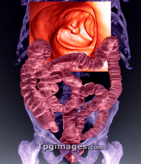
Intestines with colon polyp. At top is a coloured virtual colonoscopy (VC) image of a colon polyp in a 53-year-old patient. A VC is constructed from 3D CT (computed tomography) scans, which are entirely non-invasive. The pedunculate (stalked) polyp is seen at centre of the VC showing the colon, the lower part of the large intestine. Such polyps are benign (non-cancerous) growths that arise from the lining of the colon. Although benign, polyps are generally removed surgically as their growth may obstruct the passage of food, and some polyps do turn cancerous over time. A 3D CT scan has also been used to assess the overall structure of the intestine (pink) as it sits in the abdomen, and to check whether it has a normal position in relation to the patient's skeleton (purple).
| px | px | dpi | = | cm | x | cm | = | MB |
Details
Creative#:
TOP03215587
Source:
達志影像
Authorization Type:
RM
Release Information:
須由TPG 完整授權
Model Release:
N/A
Property Release:
N/A
Right to Privacy:
No
Same folder images:
 Loading