
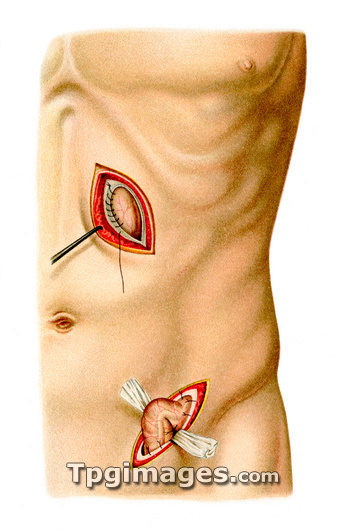
Stomach and colon surgery. Historical artwork of the techniques used in gastrostomy and colostomy. In both cases, the skin (pink), fat (yellow), muscle (red) and connective tissue membrane (white) layers have been dissected and pulled apart to reveal the abdominal organs. At top, a portion of the anterior wall of the stomach has been sutured into the wound in the abdominal wall. At bottom, a loop of the colon's sigmoid flexure has been drawn forward and fixed in the wound. These fistulas allow the patient to be fed directly into the stomach, and for faeces to be removed directly from the colon. This is usually due to disorders elsewhere in the digestive system. Artwork from Atlas and Epitome of Operative Surgery (1898, Otto Zuckerkandl).
| px | px | dpi | = | cm | x | cm | = | MB |
Details
Creative#:
TOP03220564
Source:
達志影像
Authorization Type:
RM
Release Information:
須由TPG 完整授權
Model Release:
N/A
Property Release:
N/A
Right to Privacy:
No
Same folder images:
 Loading