
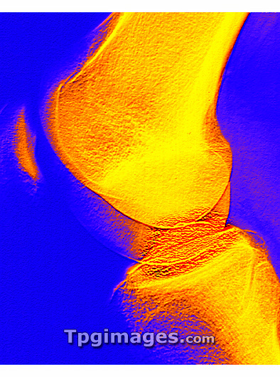
Knee joint. Coloured X-ray of a healthy human knee joint in side view, seen while bent. Two bones meet at the knee, forming a joint that works like a hinge. At top is the large femur (thigh-bone), which articulates with the tibia (shin-bone) at bottom. The patella or kneecap (upper left) is a triangular protective bone at the front of the knee held in position by muscles and tendons. Two discs of protective cartilage (not clearly visible) cover the surfaces of the femur and tibia to reduce friction between these bones. The round bottom surface of the femur forms a narrow ridge that moves through a groove in the tibia. This allows hinge movement, with slight rotation.
| px | px | dpi | = | cm | x | cm | = | MB |
Details
Creative#:
TOP03220744
Source:
達志影像
Authorization Type:
RM
Release Information:
須由TPG 完整授權
Model Release:
N/A
Property Release:
N/A
Right to Privacy:
No
Same folder images:
 Loading