
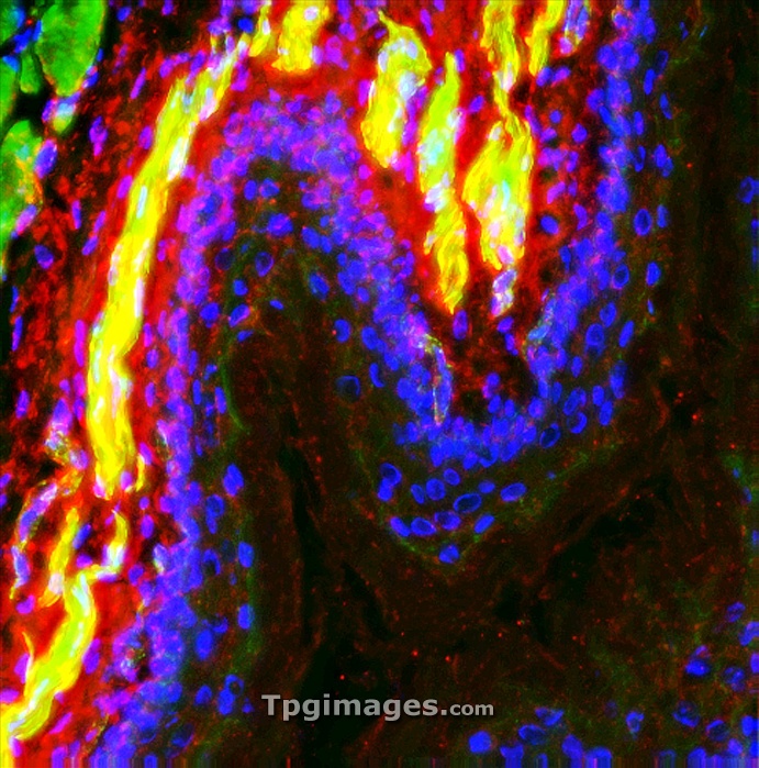
Rat oesophagus. Fluorescence deconvolution micrograph of a section through a healthy rat oesophagus (gullet), showing the co-localisation of f-actin (yellow) and smooth muscle (red) in the walls. Blue shows the cell nuclei. Magnification x50 when printed 10 centimetres wide.
| px | px | dpi | = | cm | x | cm | = | MB |
Details
Creative#:
TOP06663921
Source:
達志影像
Authorization Type:
RM
Release Information:
須由TPG 完整授權
Model Release:
NO
Property Release:
NO
Right to Privacy:
No
Same folder images:
animalanatomybiologyhistologyzoologylightmicrographfluorescentdeconvolutionmicrographconfocallightmicrographlightmicroscopeactinanatomicalbiologicalcellcellsesophagealesophagusfaunafluorescenceg-actingastrointestinalgullethealthyhistologicalhistopathologicallmmedicalmusclesmuscularnaturenormalnucleinucleusoesophagealoesophagusproteinsectionsectionedsmoothmusclestructuretissuewallwildlifezoological"
"actinanatomicalanatomyanimalbiologicalbiologycellcellsconfocaldeconvolutionesophagealesophagusfaunafluorescencefluorescentg-actingastrointestinalgullethealthyhistologicalhistologyhistopathologicallightlightlightlmmedicalmicrographmicrographmicrographmicroscopemusclemusclesmuscularnaturenormalnucleinucleusoesophagealoesophagusproteinsectionsectionedsmoothstructuretissuewallwildlifezoologicalzoology
 Loading