
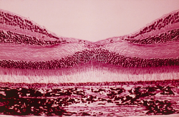
Light micrograph through a section of the human eye in the region of the fovea, a depression in the photosensitive retina, the inner lining of the eye. The foveal retina is the area of greatest visual acuity and contains only cone receptor cells (the hump-backed layers). N.B. The retina is composed of 2 types of photosensitive cells, rods & cones. The rods differentiate light of differing intensity (analagous to a black & white image); cone cells are of 3 types receptive to blue, green & red light & form a system for colour perception. The broad bottom layer in image is the choroid layer, supporting & separated from the retina by a layer of pigmented epithelial cells.
| px | px | dpi | = | cm | x | cm | = | MB |
Details
Creative#:
TOP10219980
Source:
達志影像
Authorization Type:
RM
Release Information:
須由TPG 完整授權
Model Release:
N/A
Property Release:
N/A
Right to Privacy:
No
Same folder images:
 Loading