
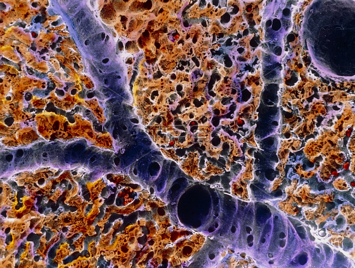
Liver tissue. False-colour scanning electron micrograph (SEM) of blood vessels around a lobule, the functional unit of the liver. A typical lobule is bound on its periphery by groups of ducts & vessels, most prominently the conducting portal vein (large blue sectioned vessel) & branches called distributing portal veins (smaller blue vessel). Plates of hepatocytes or liver cells (coloured gold) radiate towards a central vein (top right). The dark spaces surrounding them are capillaries (sinusoids). Arterial vessels also bind each lobule, but are less visible here. Magnification: x125 at 6x7cm size.
| px | px | dpi | = | cm | x | cm | = | MB |
Details
Creative#:
TOP10220706
Source:
達志影像
Authorization Type:
RM
Release Information:
須由TPG 完整授權
Model Release:
N/A
Property Release:
N/A
Right to Privacy:
No
Same folder images:
 Loading