
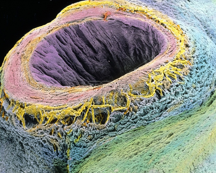
Umbilical cord. Coloured Scanning Electron Micro- graph (SEM) of a sectioned human umbilical cord. Here, the cut umbilical vein (pink) reveals its inner folded lumen and a thin inner endothelial layer (purple). Most of the vein consists of several circular layers of smooth muscle. Around it is the jelly-like mucous connective tissue (blue) known as Wharton's jelly. Not seen are the two umbilical arteries which spiral around the vein. The umbilical cord is the attachment connecting the foetus with the placenta, and by which the foetus receives nourishment from the mother's blood. Magnification: x20 at 6x7cm size. x26 at 4x5"~LANDSCAPE"
| px | px | dpi | = | cm | x | cm | = | MB |
Details
Creative#:
TOP10221389
Source:
達志影像
Authorization Type:
RM
Release Information:
須由TPG 完整授權
Model Release:
N/A
Property Release:
N/A
Right to Privacy:
No
Same folder images:
 Loading