
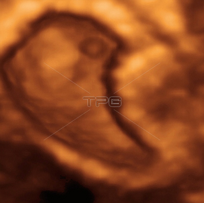
Embryo. Coloured 3-D ultrasound scan of a six- week-old embryo in the uterus. At this stage of development the embryo measures about 13 millimetres in length but has no identifiable structures. It is known as the foetal pole. The spine and spinal cord are growing rapidly at this stage. Ultrasound scanning is a diagnostic technique that sends high-frequency sound waves into the body via a transducer. The returning echoes are recorded and used to build an image of an internal structure. Foetal ultrasound scanning is routine during pregnancy. 3-D scanning uses computer technology to return more detailed images than conventional 2-D scans.
| px | px | dpi | = | cm | x | cm | = | MB |
Details
Creative#:
TOP10222420
Source:
達志影像
Authorization Type:
RM
Release Information:
須由TPG 完整授權
Model Release:
N/A
Property Release:
N/A
Right to Privacy:
No
Same folder images:
 Loading