
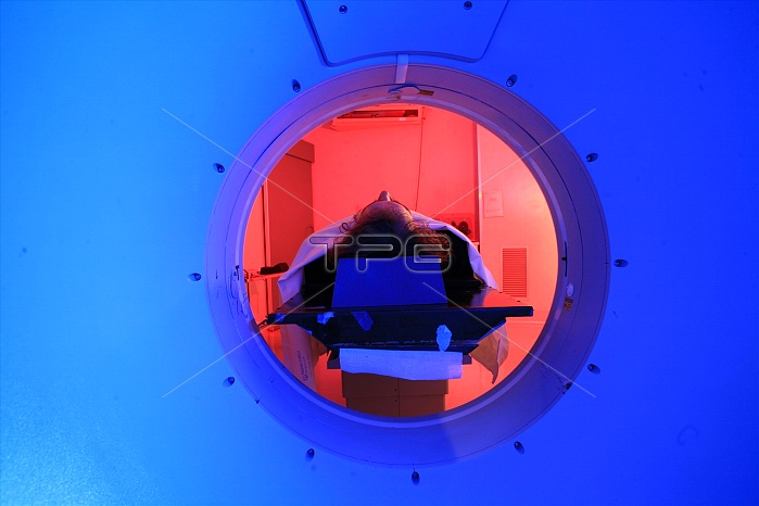
PET: Positron emission tomography is a nuclear medicine imaging technique which produces a three dimensional image or picture of functional processes in the body. The system detects pairs of gamma rays emitted indirectly by a positron emitting radionuclide (tracer), which is introduced into the body on a biologically active molecule. Images of tracer concentration in three dimensional space within the body are then reconstructed by computer analysis. In modern scanners, this reconstruction is often accomplished with the aid of a CT X ray scan performed on the patient during the same session, in the same machine. PET is both a medical and research tool. It is used heavily in clinical oncology ( medical imaging of tumors and the search for metastases), and for clinical diagnosis of certain diffuse brain diseases such as those causing various types of dementias. PET is also an important research tool to map normal human brain and heart function. Provides metabolic images, since the uptake and the distribution of the radipharmaceutical depends on tumour metabolism and the background activity. The PET unit of The IRCCS is involved in the production of radioisotopes by cyclotron. The pet unit is equipped to study patients with virtually all types of human malignancies for staging and follow up.
| px | px | dpi | = | cm | x | cm | = | MB |
Details
Creative#:
TOP11657197
Source:
達志影像
Authorization Type:
RM
Release Information:
須由TPG 完整授權
Model Release:
No
Property Release:
No
Right to Privacy:
No
Same folder images:
 Loading