
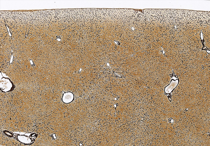
Light microscopy of liver tissue using the reticulin stain whereby silver is deposited on collagen fibres. The liver cells (orange-brown hepatocytes) are stained with neutral red stain. The larger diameter holes are branches of blood vessels bringing blood into the liver. The reticulin method also reveals the many small blood vessels (called vascular sinusoids) forming radial patterns in the form of indistinct polygons with empty, small blood vessels (called central veins) at the centre. These represent the hepatic lobules through which blood percolates among the hepatocytes. Blood leaves a lobule by passing into the central veins that ultimately unite to form hepatic veins passing to the inferior vena cava. Magnification x25 when printed at 10 cm.
| px | px | dpi | = | cm | x | cm | = | MB |
Details
Creative#:
TOP14988168
Source:
達志影像
Authorization Type:
RM
Release Information:
須由TPG 完整授權
Model Release:
N/A
Property Release:
No
Right to Privacy:
No
Same folder images:
 Loading