
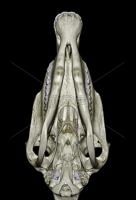
Underside of a horse's skull. 3D computed tomography (CT) scan of the skull of a horse (Equus caballus), seen from underneath. The front of the skull (and the mouth) is at top. At top centre are the incisor teeth. Behind these is the interdental space, with the premolar and molar teeth at left and right. The eye sockets are at lower left and lower right, next to the temporomandibular joints (TMJ). For this skull as a 3D CT scan from the side, see image C025/4075. For this skull showing the pulp of the teeth, see image C025/4076.
| px | px | dpi | = | cm | x | cm | = | MB |
Details
Creative#:
TOP15316770
Source:
達志影像
Authorization Type:
RM
Release Information:
須由TPG 完整授權
Model Release:
N/A
Property Release:
N/A
Right to Privacy:
No
Same folder images:
3-d3danatomicalanatomyanimalbiologicalbiologyblackbackgroundbonebonybonyorbitcomputedtomographycraniumctscannercutoutcutoutscut-outcut-outscutoutcutoutsequineequuscaballuseyesocketeyesocketsfaunahealthyhorseincisorincisorsinferiorviewinterdentalspacejawjointlowerjawmammalmandiblemaxillamolarmolarsnaturenormalpremolarpremolarsskullspecimenteethtemporalbonetemporomandibularjointthreedimensionaltmjtoothunderneathupperjawviewfrombelowwildlifezoologicalzoology
3-d3danatomicalanatomyanimalbackgroundbelowbiologicalbiologyblackbonebonebonybonycaballuscomputedcraniumctcutcutcut-outcut-outscutoutcutoutsdimensionalequineequuseyeeyefaunafromhealthyhorseincisorincisorsinferiorinterdentaljawjawjawjointjointlowermammalmandiblemaxillamolarmolarsnaturenormalorbitoutoutspremolarpremolarsscannerskullsocketsocketsspacespecimenteethtemporaltemporomandibularthreetmjtomographytoothunderneathupperviewviewwildlifezoologicalzoology
 Loading