
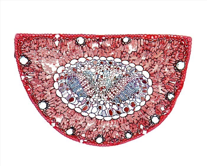
Scots pine (Pinus sylvestris) leaf. Light micrograph of a section through a needle (leaf) from a Scots pine tree, showing its thick cuticle and thick-walled epidermis (red) and hypodermis (subcutaneous tissue, brown). Stomata (pores) and guard cells can also be seen, along with chloroplasts, and mesophyll tissue composed of parenchyma cells. The vascular tissue is composed of xylem tracheid cells (red) and phloem sieve tube cells (blue). Magnification: x37 when printed 10 centimetres wide.
| px | px | dpi | = | cm | x | cm | = | MB |
Details
Creative#:
TOP16630766
Source:
達志影像
Authorization Type:
RM
Release Information:
須由TPG 完整授權
Model Release:
N/A
Property Release:
N/A
Right to Privacy:
No
Same folder images:
anatomicalanatomybiologicalbiologybotanicalbotanycellcellschloroplastchloroplastsconiferconiferouscutoutcutoutscut-outcut-outscutoutcutoutsepidermalepidermisfloraguardcellgymnospermgymnospermshistologicalhistologyhypodermisleaflightmicrographlightmicroscopemesophyllnatureneedleno-onenobodyparenchymaphloempinussylvestrisplantplantsporescotspinesectionsectionedsievetubesievetubesstomastomatasubcutaneoustissuetracheidtracheidstreevascularbundlewhitebackgroundwildlifexerophyticxylem
anatomicalanatomybackgroundbiologicalbiologybotanicalbotanybundlecellcellcellschloroplastchloroplastsconiferconiferouscutcutcut-outcut-outscutoutcutoutsepidermalepidermisfloraguardgymnospermgymnospermshistologicalhistologyhypodermisleaflightlightmesophyllmicrographmicroscopenatureneedleno-onenobodyoutoutsparenchymaphloempinepinusplantplantsporescotssectionsectionedsievesievestomastomatasubcutaneoussylvestristissuetracheidtracheidstreetubetubesvascularwhitewildlifexerophyticxylem
 Loading