
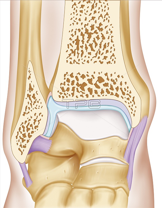
Anterior view illustration of the ankle joint. The two joint surfaces (grey) are visible. the joint surface of the fibula and tibia distal epiphysis and that of the talus. The articular capsule is filled with synovial fluid (blue) surrounded by its membrane (pink). it is attached to the lateral ligaments.
| px | px | dpi | = | cm | x | cm | = | MB |
Details
Creative#:
TOP18617124
Source:
達志影像
Authorization Type:
RM
Release Information:
須由TPG 完整授權
Model Release:
No
Property Release:
No
Right to Privacy:
No
Same folder images:
 Loading