
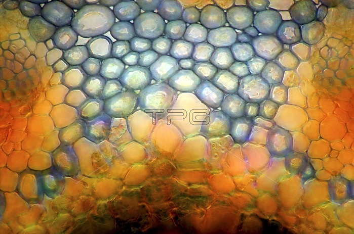
Pine needle. Darkfield illuminated polarised light micrograph of a cross-section through a pine needle (Pinus sp.). The section shows transfusion tissue within the endodermis in the centre of the needle. Transfusion tissue is composed of parenchyma cells (blue) which fill the space between the vascular bundles (left and right). Magnification: x400, when printed at 10 centimetres wide.
| px | px | dpi | = | cm | x | cm | = | MB |
Details
Creative#:
TOP19257365
Source:
達志影像
Authorization Type:
RM
Release Information:
須由TPG 完整授權
Model Release:
N/A
Property Release:
N/A
Right to Privacy:
No
Same folder images:
anatomicalanatomybiologicalbiologybotanicalbotanybundlecellcellsconiferconiferouscross-sectiondarkfielddarkfieldendodermisevergreenfloragymnospermgymnospermshistologicalhistologyleaflightlightmicrographlightmicroscopelmmicroscopemicroscopicnatureneedleno-onenobodyparenchymapinepineleafpineneedlepinuspinusleafpinusneedleplantpolarisedpolarizedtissuetransfusiontissuetransversesectiontreevascularvascularbundle
anatomicalanatomybiologicalbiologybotanicalbotanybundlebundlecellcellsconiferconiferouscross-sectiondarkdarkfieldendodermisevergreenfieldfloragymnospermgymnospermshistologicalhistologyleafleafleaflightlightlightlmmicrographmicroscopemicroscopemicroscopicnatureneedleneedleneedleno-onenobodyparenchymapinepinepinepinuspinuspinusplantpolarisedpolarizedsectiontissuetissuetransfusiontransversetreevascularvascular
 Loading