
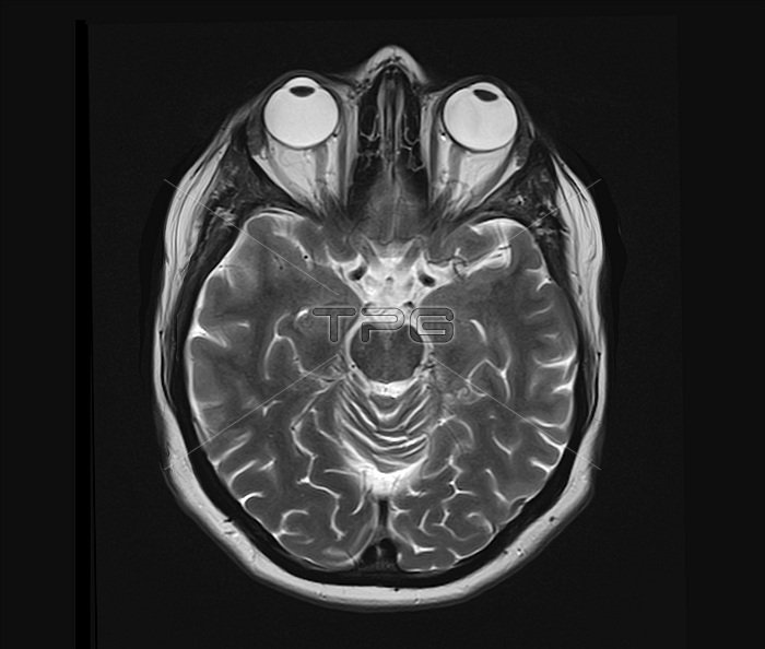
Human eyes and brain. Axial magnetic resonance imaging (MRI) scan of the eyes (top) and brain of a 33-year-old man. The scan shows the normal structures, including the eyeballs and the optic nerves and the crossing point of the optic nerves (the optic chiasma). The optic nerves run back from the eyes and cross at a point behind the nose, which is seen between the eyes. Structures separating the two brain hemispheres are also seen. This scan passes through a reference plane that is called the neuro-ocular plane (NOP). This MRI scan is an example of a T2-weighted scan, with the use of gadolinium as a contrast medium.
| px | px | dpi | = | cm | x | cm | = | MB |
Details
Creative#:
TOP19633685
Source:
達志影像
Authorization Type:
RM
Release Information:
須由TPG 完整授權
Model Release:
N/A
Property Release:
N/A
Right to Privacy:
No
Same folder images:
 Loading