
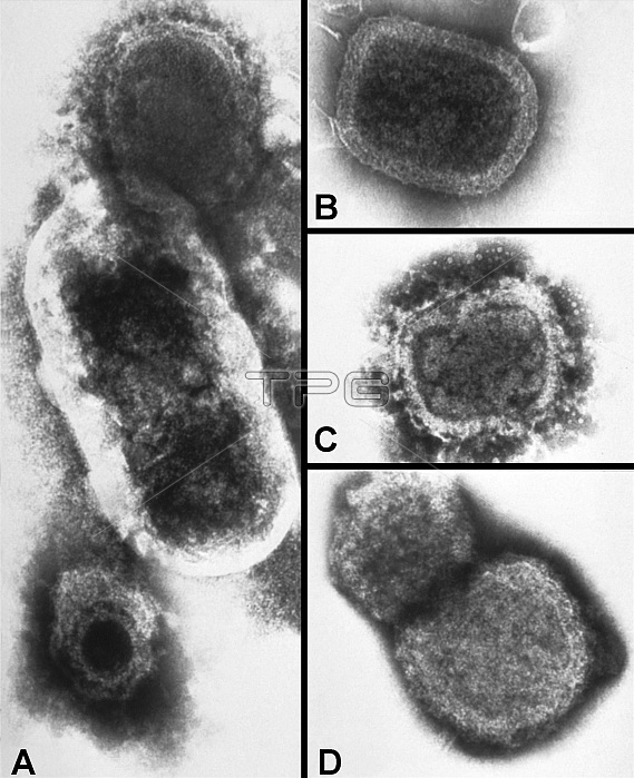
Negatively-stained transmission electron microscopic (TEM) images revealed some of the ultrastructural morphology exhibited by a number of different microorganisms. Panel "A" represents a composite micrograph, created for the purpose of comparing the size difference between a poxvirus at the top, a bacillus in the middle, and a herpesvirus at the bottom. Panels "B", "C", and "D" are TEMs depicting the sequential degeneration of variola virus particles.
| px | px | dpi | = | cm | x | cm | = | MB |
Details
Creative#:
TOP22238059
Source:
達志影像
Authorization Type:
RM
Release Information:
須由TPG 完整授權
Model Release:
N/A
Property Release:
No
Right to Privacy:
No
Same folder images:
bwdegenerationtemmicrographytransmissionelectronmicroscopytransmissionelectronmicrographemelectronmicroscopyelectronmicrographmicroscopymicroscopicmicrographdumbbell-shapedbrick-shapedviraldnaviraldiseaseinfectiousdiseaseinfectioncontagiousdiseaseacutediseasebacteriologybacteriumbacterialbacteriabacillusherpesviralesherpesviridaeherpesvirusherpesvirusherpespoxviruspoxviridaeorthopoxorthopoxvirusvariolaveravariolasmallpoxpathogenicpathogenpathologicalpathologyvirologyvirusparticlesvirionviralvirusultrastructuralmorphologymicroscopicorganismmicroorganismmicrobiologyscience
acutebacillusbacteriabacterialbacteriologybacteriumbrick-shapedbwcontagiousdegenerationdiseasediseasediseasediseasednadumbbell-shapedelectronelectronelectronelectronemherpesherpesherpesviralesherpesviridaeherpesvirusinfectioninfectiousmicrobiologymicrographmicrographmicrographmicrographymicroorganismmicroscopicmicroscopicmicroscopymicroscopymicroscopymorphologyorganismorthopoxorthopoxvirusparticlespathogenpathogenicpathologicalpathologypoxviridaepoxvirussciencesmallpoxtemtransmissiontransmissionultrastructuralvariolavariolaveraviralviralviralvirionvirologyvirusvirusvirus
 Loading