
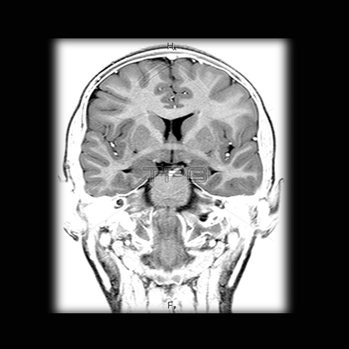
This coronal (frontal view) T1 weighted MRI image of the brain demonstrates normal cerebral anatomy. In this view you see the anterior commisure, which is a white matter tract connecting the temporal lobes. Also seen in this image is the amygdala, located along the medial temporal lobes, just anterior and superior to the head of the hippocampus.
| px | px | dpi | = | cm | x | cm | = | MB |
Details
Creative#:
TOP22291097
Source:
達志影像
Authorization Type:
RM
Release Information:
須由TPG 完整授權
Model Release:
N/A
Property Release:
No
Right to Privacy:
No
Same folder images:
 Loading