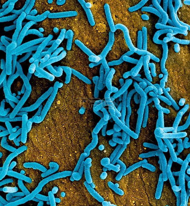
Colorized scanning electron micrograph of Marburg virus particles (blue) both budding and attached to the surface of infected VERO E6 cells (orange). Image captured and color-enhanced at the NIAID Integrated Research Facility in Fort Detrick, Maryland. Credit: NIAID.
| px | px | dpi | = | cm | x | cm | = | MB |
Details
Creative#:
TOP26089420
Source:
達志影像
Authorization Type:
RM
Release Information:
須由TPG 完整授權
Model Release:
N/A
Property Release:
N/A
Right to Privacy:
No
Same folder images:
AmericaNorthAmericaUnitedStatesMarylandanimalcatchorangebluesurfaceparticlesscientificimagerymedicalimagerymicroscopeelectronicmicroscopeSEMinfectionreproductivesystembuddingcytologycellcellculturecelllineimmortalizedcelllineVeroE6cellresearchcentrevirusRNAvirusfiloviridaefilovirusmarburgvirusmarburgviruscolorization
AmericaAmericaE6MarylandNorthRNASEMStatesUnitedVeroanimalbluebuddingcatchcellcellcellcellcellcentrecolorizationculturecytologyelectronicfiloviridaefilovirusimageryimageryimmortalizedinfectionlinelinemarburgmarburgvirusmedicalmicroscopemicroscopeorangeparticlesreproductiveresearchscientificsurfacesystemvirusvirusvirus

 Loading