
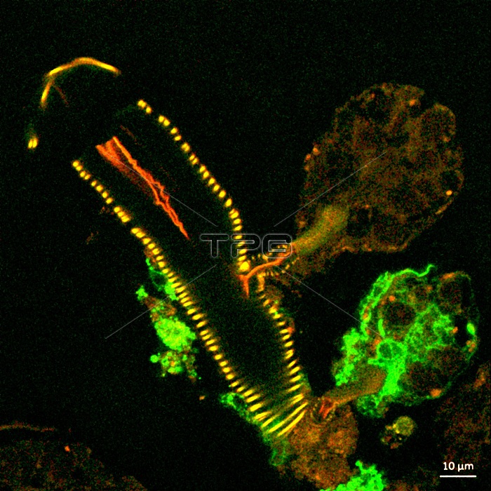
This confocal microscope image shows a cross section of a tick salivary gland infected with Langat virus (green). Two rounded structures on the right, called acini, are shown attached to a duct (yellow). The lower acinus is infected, as denoted by the green fluorescent signal. Credit: NIAID.
| px | px | dpi | = | cm | x | cm | = | MB |
Details
Creative#:
TOP26089523
Source:
達志影像
Authorization Type:
RM
Release Information:
須由TPG 完整授權
Model Release:
N/A
Property Release:
N/A
Right to Privacy:
No
Same folder images:
 Loading