
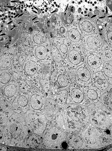
Transmission electron micrograph (TEM) of the ultrastructure of the seminiferous epithelium in the testis. Adjacent to the basal lamina is a Sertoli cell nucleus (centrally) and spermatogonia. Above these are primary spermatocytes with condensed chromatin in their nuclei, and numerous round spermatids some of which show linkage by intercellular cytoplasmic bridges. Compacted slender elongated sperm heads are close to the lumen of the seminiferous tubule. At this position are cross-sectioned sperm tails and the discarded cytoplasm of spermatids termed residual bodies or residual cytoplasm. Thin branches of Sertoli cell cytoplasm (similar to tree branches) extend between and support the germ cells. Magnification: x1,000 when width printed at 10cm.
| px | px | dpi | = | cm | x | cm | = | MB |
Details
Creative#:
TOP27945067
Source:
達志影像
Authorization Type:
RM
Release Information:
須由TPG 完整授權
Model Release:
N/A
Property Release:
N/A
Right to Privacy:
No
Same folder images:
 Loading