
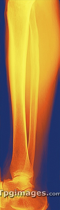
Normal lower leg, coloured frontal X-ray. The tibia (shinbone, left) and the smaller fibula (centre) articulate with the foot bones (bottom) to form the ankle joint. The outline of the calf muscle is seen at upper right.
| px | px | dpi | = | cm | x | cm | = | MB |
Details
Creative#:
TPG05326249
Source:
達志影像
Authorization Type:
RF
Release Information:
須由TPG 完整授權
Model Release:
NO
Property Release:
NO
Right to Privacy:
No
Same folder images:
 Loading