
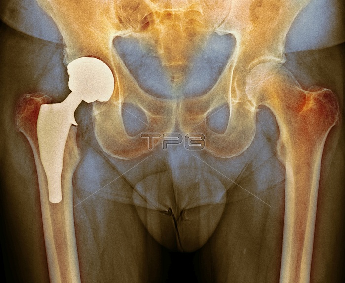
Total hip replacement. Coloured X-ray of a section through the pelvic region of a 66-year-old male patient after total hip replacement surgery. Here, the prosthetic hip joint (light, left) can be seen, with the peg implanted in the femur (centre-left) and replacement socket in the hip (upper left).
| px | px | dpi | = | cm | x | cm | = | MB |
Details
Creative#:
TPG20298660
Source:
達志影像
Authorization Type:
RF
Release Information:
須由TPG 完整授權
Model Release:
N/A
Property Release:
N/A
Right to Privacy:
No
Same folder images:
NOBODYNO-ONEMEDICINEMEDICALHEALTHCAREABNORMALUNHEALTHYCONDITIONDISORDERDISEASEDISEASEDSECTIONSECTIONEDMALEMAN6666-YEAR-OLD66-YEARS-OLDSIXTIESX-RAYX-RAYSXRAYXRAYSDIAGNOSTICIMAGINGRADIOLOGYRADIOLOGICALRADIOGRAPHYRHEUMATOLOGYRHEUMATOLOGICALORTHOPAEDICORTHOPAEDICSORTHOPEDICORTHOPEDICSBONEBONESJOINTCOLOUREDCOLOREDFALSE-COLOREDFALSE-COLOUREDTOTALHIPREPLACEMENTTHRPELVISPELVICFEMURIMPLANTIMPLANTEDPROSTHETICPROSTHESISPROSTHESESBALLANDSOCKETPOST-OPPOST-OPERATIVESURGICALSURGERYOPERATIONAFTERIMPLANTED
6666-YEAR-OLD66-YEARS-OLDABNORMALAFTERANDBALLBONEBONESCOLOREDCOLOUREDCONDITIONDIAGNOSTICDISEASEDISEASEDDISORDERFALSE-COLOREDFALSE-COLOUREDFEMURHEALTHCAREHIPIMAGINGIMPLANTIMPLANTEDIMPLANTEDJOINTMALEMANMEDICALMEDICINENO-ONENOBODYOPERATIONORTHOPAEDICORTHOPAEDICSORTHOPEDICORTHOPEDICSPELVICPELVISPOST-OPPOST-OPERATIVEPROSTHESESPROSTHESISPROSTHETICRADIOGRAPHYRADIOLOGICALRADIOLOGYREPLACEMENTRHEUMATOLOGICALRHEUMATOLOGYSECTIONSECTIONEDSIXTIESSOCKETSURGERYSURGICALTHRTOTALUNHEALTHYX-RAYX-RAYSXRAYXRAYS
 Loading