
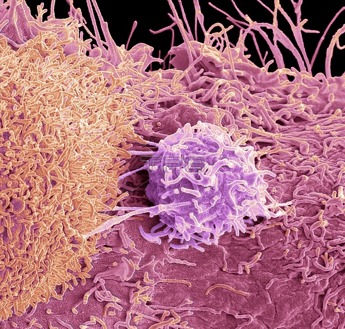
Prostate cancer cell, coloured scanning electron micrograph (SEM). of the surface of prostate cancer cells. The cells shows numerous processes and microvilli (fine surface projections). These features are characteristic of highly mobile cells, and enable cancerous cells to spread (metastasis) rapidly round the body, and invade other organs and tissues. Cancer cells divide rapidly and chaotically, and may clump to form malignant tumours. The prostate is a small gland found in men just below the bladder, surrounding the urethra, the tube urine passes through. Prostate cancer is most prevalent in men over 50 years of age. Treatment is with hormone therapy, chemotherapy, or surgical removal of the prostate. Magnification: x 8000 when printed at 10 centimetres across.
| px | px | dpi | = | cm | x | cm | = | MB |
Details
Creative#:
TPG30861446
Source:
達志影像
Authorization Type:
RF
Release Information:
須由TPG 完整授權
Model Release:
N/A
Property Release:
N/A
Right to Privacy:
No
Same folder images:
 Loading